File:Fig4 Rubel FInNeuroinformatics2016 10.jpg

Original file (964 × 570 pixels, file size: 257 KB, MIME type: image/jpeg)
Summary
| Description |
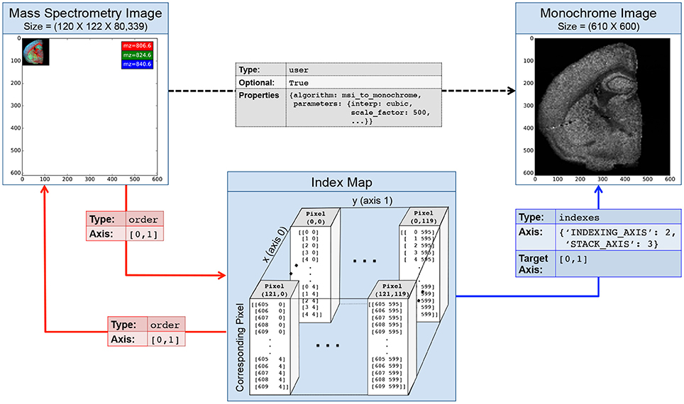
Figure 4. Illustration of an index map relationship describing the interaction between a mass spectrometry image (MSI) and a derived monochrome image. The MSI image is in this case 5× smaller than the monochrome image. The intermediary index map describes for each pixel in the MSI image which pixels it corresponds to in the the monochrome image. Two order relationships (red arrows) describe the interactions between the MSI image and the map and vice versa. A third indexes relationship links our index map to the monochrome image and describes how the map can be used to access the image. Optionally, we may create a fourth user relationship (black arrow) to further characterize the semantic relationship between the derived and original image (e.g., to store a description of the algorithm and parameters used to generate the image). Naturally, we can also describe the inverse mapping between the original and processed image via a second index map relationship. |
|---|---|
| Source |
Rübel, O.; Dougherty, M.; Prabhat; Denes, P.; Conant, D.; Chang, E.F.; Bouchard, K. (2016). "Methods for specifying scientific data standards and modeling relationships with applications to neuroscience". Frontiers in Neuroinformatics 10: 48. doi:10.3389/fninf.2016.00048. PMID 27867355. |
| Date |
2016 |
| Author |
Rübel, O.; Dougherty, M.; Prabhat; Denes, P.; Conant, D.; Chang, E.F.; Bouchard, K. |
| Permission (Reusing this file) |
|
| Other versions |
Licensing
|
|
This work is licensed under the Creative Commons Attribution 4.0 License. |
File history
Click on a date/time to view the file as it appeared at that time.
| Date/Time | Thumbnail | Dimensions | User | Comment | |
|---|---|---|---|---|---|
| current | 21:44, 20 February 2017 |  | 964 × 570 (257 KB) | Shawndouglas (talk | contribs) |
You cannot overwrite this file.
File usage
The following page uses this file:


